| 08:30 to 09:00 |
Registration
|
| 09:00 to 09:10 |
President's Introduction
|
| 09:09 |
SESSION ONE - Moderator: Cemal Ucer, ADI President
|
| 09:10 to 09:30 |
Improving conventional periodontal subepithelial connective tissue grafting for the demands of peri-implantology Following tooth extraction, resorption of the buccal wall of the socket will occur; this is true for both the maxilla and mandible. Where the extraction site is surrounded by natural dentition, the loss of the buccal alveolar wall can degrade the visual aesthetics of an implant-supported prosthetic rehabilitation.
To aid harmonization of hard and soft tissue morphology, both hard and soft tissue augmentation can be carried out either consecutively with an extraction/immediate implant placement or prior to an implant placement in the delayed scenario. The contemporary method of increasing soft tissue volume is to use the subepithelial connective tissue (auto) graft (the SCTG). The graft requires fixation, otherwise it can be extruded from the recipient site.
This presentation will be a step-by-step description of a collection of innovatory suturing techniques developed by the author to be specifically applied to dental implantology, for the preparation and improvement of dental implant sites; all can confidently secure the SCTG, thus resisting its dislodgement. The presentation will be illustrated by (at least) 20 cases with follow-ups ranging from a minimum of 12 months to 60 months.
Shane McCrea 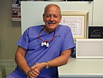 Shane McCrea graduated from the Royal Dental Hospital, London University in 1979, becoming a member of the University of Dusseldorf's Work Study Groups in implantology, prosthetics and periodontology until 2001. Shane McCrea graduated from the Royal Dental Hospital, London University in 1979, becoming a member of the University of Dusseldorf's Work Study Groups in implantology, prosthetics and periodontology until 2001.
He gained the MFGDP examination in 2003. In 2005 he completed an MSc in Dental and Maxillofacial Radiology (Kings College, London), his research project being published in the BDJ. As a result of this project, he is now a Peer Reviewer for fourteen dental journals.
Between 2008 and 2012 he has had seventeen papers published in peer-reviewed journals. In 2010 he gained an MMedSci (Dental Implantology) from The University of Sheffield. He is presently undertaking a further MSc in Stem Cells and Regeneration at The University of Bristol. This year, he has had three papers accepted for publication. Of the eleven case studies published by the British Society of Periodontology, four are attributed to Shane's work on soft and hard tissue management. He is now Co-ordinator of Post-Graduate Education for the British Society of Oral Implantology.
Qualifications: MMedSci (Dental Implantology) MSc (Dental and Maxillofacial Radiology) BDS (Lond) LDS RCS (Eng) MFGDP (UK)
Qualifications: MMedSci (Dental Implantology) MSc (Dental and Maxillofacial Radiology) BDS (Lond) LDS RCS (Eng) MFGDP (UK) |
| 09:30 to 09:50 |
The slow drilling protocol for implant site preparation Implant preparation is characterised by the creation of a tunnel within the alveolar bone which is used to accommodate the titanium fixture. The standard preparation of the osteotomy site with rotational drills is characterised by the conversion of mechanical energy into thermal energy. This can create a transient and localised increase in temperature above the normal physiological limits with resultant osteonecrosis and/or impairment of osteogenic potential.
A multitude of temperature abatement strategies aimed at minimising the collateral damage resulting from implant preparation are implemented and considered routine. Broadly speaking these can be classified as equipment or technique protocols. Some of the equipment protocols include the use of cooled irrigants, the deployment of sharp (or disposable) sequential drills, the use of piezo surgery. Standard technique protocols advocate the use of light pressure, a pecking motion and drill speeds of the order of 450-1000 rpm.
The slow drilling protocol utilises a significantly lower drilling speed of 150 rpm to bore through the cortical plate and 50 rpm to prepare the medullary bone. This maintains the heat generated to levels well below the physiological level for which osteonecrosis may occur. The protocol is a simple adaptation of current standard practice but confers additional advantages. It allows for the use of rotational ridge expanders, the simple and safe elevation of the sinus floor with the internal sinus lift technique, it increases significantly the quantities of autogenous bone collected and it allows for excellent integration with blood plasma technologies.
Nicholas Claydon 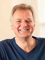 Nicholas Claydon qualified in 1986 from Cardiff Dental School and completed his foundation training to Registrar level. He attained his MScD by research at the University of Wales and his PhD from the University of Bristol. Nicholas Claydon qualified in 1986 from Cardiff Dental School and completed his foundation training to Registrar level. He attained his MScD by research at the University of Wales and his PhD from the University of Bristol.
An accredited specialist in Periodontics, Nick has established a reputation for aesthetic treatment of complex restorative cases involving periodontal, implant and prosthodontic disciplines.
He is a researcher and lecturer at the Bristol Dental School, utilising his skills as mentor for dentists managing implant cases. He is a reviewer for international research journals and conducts clinical studies which are published in peer reviewed journals. In 2009 Nick established his state of the art specialist Dental Practice at The Pines in Whitchurch.
Qualifications: BDS MScD PhD |
| 09:50 to 10:10 |
Immediate placement and immediate loading of implants placed into fresh extraction sockets Immediate placement and immediate loading of implants carries a tremendous attraction because of an inherent reduction in treatment time and considerable convenience in comparison to conventional implant treatment. The goal of this study was to evaluate the clinical outcome of implants placed immediately into fresh extraction sockets and loaded immediately.
Materials and methods: Patients were considered for immediate placement after careful assessment of the failing tooth and future implant site. A detailed protocol was followed to enable a possible immediate loading of the implant with the use of prefabricated angulated abutments (0° - 37.5°) in combination with transitional restorations.
Results: As of May 2013 a total of 843 immediate implants had been placed in 400 patients and loaded immediately. The maximum observation period by May 2013 was 162 months with a mean observation period of 39 months. Six implants had been lost in function. A broad range of prefabricated angulated abutments was required in order to be able to restore the implants immediately. Angles beyond 15° were necessary in more than 50% of the cases.
Conclusion: The preliminary results of this ongoing prospective clinical study are very promising. Immediate loading of immediately placed implants is a possible treatment option that might be predictably and successfully achieved. A simple restorative protocol for chair-side fabrication of temporary restorations can be followed in order to load the implant immediately, when a broad range of prefabricated angulated abutments (0° - 37.5°) is used.
Ashok Sethi 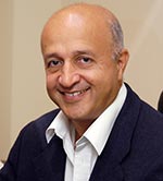 Ashok Sethi is a registered specialist in Oral Surgery as well as Prosthodontics and his Harley Street referral practice in the heart of London is dedicated solely to Implant Dentistry. Ashok Sethi is a registered specialist in Oral Surgery as well as Prosthodontics and his Harley Street referral practice in the heart of London is dedicated solely to Implant Dentistry.
He conceived and has run the Diploma in Implant Dentistry at the Royal College of Surgeons of England.
The second edition of his book ‘Practical Implant Dentistry – the Science and Art’ (co-author Thomas Kaus) has just been published by Quintessence.
Qualifications: BDS DGDP (UK) MGDSRCS (Eng) DUI (Lille) FFGDP (UK) |
| 10:10 to 10:40 |
Panel Discussion
Cemal Ucer - Moderator; Shane McCrea - Dorset; Nicholas Claydon - Wales; Ashok Sethi - London
|
| 10:40 to 11:00 |
Tea/Coffee & Exhibition
|
| 11:00 to 11:20 |
My biggest implant disaster: How it happened, how I managed it, and how you can avoid it happening to you If you're the kind of clinician who never makes a mistake, then this presentation is not for you - you won't learn anything useful from it and your time is better spent elsewhere.
Like most clinicians, however, I do make mistakes and here I will present a clinical case that became my biggest implant disaster. A simple, stupid mistake on my part turned into a nightmare for both the patient and me - though she suffered much, much more as a result.
I will explain the simple error that led to my negligence (for that's what this was), and how the case quickly spiralled from a relatively localised problem into a larger and larger disaster.
I will explain how I managed the case - and I do not claim that this management was in any way ideal - and discuss what I might have done better.
Finally, I will explain how some simple steps can help you can avoid the same thing happening to you and your patients.
Bill Schaeffer 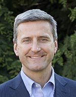 Bill Schaeffer is qualified in dentistry and medicine, has postgraduate qualifications in both dental and general surgery and is recognised as a specialist oral surgeon. He has been placing dental implants for more than 16 years and is experienced in many different implant systems. Bill Schaeffer is qualified in dentistry and medicine, has postgraduate qualifications in both dental and general surgery and is recognised as a specialist oral surgeon. He has been placing dental implants for more than 16 years and is experienced in many different implant systems.
Bill works full-time doing implant surgery in Sussex and has lectured widely on the subject both in the UK and abroad.
Qualifications: BDS MBBS FDSRCS (Eng) MRCS (Eng) |
| 11:20 to 11:40 |
Ridge preservation in the lower molar area The loss of a tooth especially when associated with extensive pathology in the lower molar area may often lead to one of the most difficult situations in implant placement.
When allowing for full healing of 3 to 4 months the modelling of the hard tissue is often mostly on the buccal aspect which can lead to angled bone profile complicating implant placement at that time. But it may also lead to the loss of the attached gingival tissue and this can have a detrimental effect on the long term health of the Implant.
Thus ridge preservation may be of great value in reducing this early bone loss with the associated retention of the attached keratinised soft tissue.
This can be achieved in two ways, firstly using delayed immediate placement at 3 weeks post extraction where we have soft tissue closure but prior to hard tissue modelling (Schropp). Secondly where the socket is devoid of good hard tissue or vital tissues (IAN) and areas (Sinus) are too close, preventing placement. Here we can socket graft and place later at 3-4 months.
Peter Fairbairn  Peter Fairbairn is currently visiting Professor in the Department of Periodontology and Implant Dentistry at the University of Detroit Mercy School of Dentistry, Michigan, US and has been involved in the placement of Dental Implants since 1991. He was initially taught and mentored by Barry Edwards and for the last 10 years has solely used synthetic particulate graft materials in the regeneration of bone. Peter Fairbairn is currently visiting Professor in the Department of Periodontology and Implant Dentistry at the University of Detroit Mercy School of Dentistry, Michigan, US and has been involved in the placement of Dental Implants since 1991. He was initially taught and mentored by Barry Edwards and for the last 10 years has solely used synthetic particulate graft materials in the regeneration of bone.
With nearly 2,000 grafts in this period Peter has been able to develop not only materials but also the techniques in their use to hopefully improve the outcomes for the Dentist and Patient alike.
He is an active member of the ADI, BACD, BDA and the Honorary Secretary of the London Dental Fellowship.
Qualifications: BDS (Rand)
|
| 11:40 to 12:00 |
The bone ring technique: New perspectives in augmentation A two-stage method is usually chosen for bone defect augmentations with autogenous blocks and then restored by implant placement.
An alternative procedure is a bone ring technique that allows bone transplantation and implantation to be performed on large three-dimensional bone defects in a single operation.
The technique is applicable for almost all indications, including a sinus lift with minimal bone height. In cases of large bone defects or advanced jaw atrophy, extensive horizontal and/or vertical bone augmentation is required to prepare the patient’s implant treatment. This traditionally occurs in a time-consuming, painful and risky procedure – by harvesting and transplantation of the patient’s own autologous bone in a two-stage procedure (augmentation/implantation).
The bone ring technique is performed using pre-fabricated cancellous ring-shaped allograft of processed allogenic donor bone, which is placed by press-fit into a trephine drill-prepared bone bed. At the same time an implant is inserted into the ring. The bony integration of both the bone ring and the implant occurs via the surrounding vital bone. The allograft bone ring technique allows bone augmentation and implantation in a one-stage procedure.
Bone ring eliminates the need for autologous bone harvesting and manual adaptation of autologous blocks. Thereby, allograft bone ring reduces pain, risk of infection, morbidity, operation time and total procedure costs significantly. The bone ring allows for both vertical and horizontal augmentation and new bone formation, therefore simplifying the surgical treatment of three dimensional bone defects. The entire patient’s treatment period is shortened by several months and saves re-entry procedures.
Orcan Yüksel  Orcan Yüksel graduated from Johan Wolfgang Goethe University in Frankfurt am Main and Istanbul University in 1987, and subsequently took a doctor degree. Since 1993 he has owned a dental clinic with education center, specialized in dental aesthetics, oral implantology and adhesive dentistry.
He is running a number of education programs as a certified implantologist and implantological trainer of European Association of Dental Implantology (BDIZ/EDI) and Diplomate of ICOI, is a guest lecturer in the certified Curriculum Program of Oral Implantology and accredited supervisor in Master of Oral Implantology at Frankfurt University. He is a tutor in P3 (Personal Performance Program for young implantologists, future international speakers) groups’ development. Orcan Yüksel graduated from Johan Wolfgang Goethe University in Frankfurt am Main and Istanbul University in 1987, and subsequently took a doctor degree. Since 1993 he has owned a dental clinic with education center, specialized in dental aesthetics, oral implantology and adhesive dentistry.
He is running a number of education programs as a certified implantologist and implantological trainer of European Association of Dental Implantology (BDIZ/EDI) and Diplomate of ICOI, is a guest lecturer in the certified Curriculum Program of Oral Implantology and accredited supervisor in Master of Oral Implantology at Frankfurt University. He is a tutor in P3 (Personal Performance Program for young implantologists, future international speakers) groups’ development.
He has published numerous international presentations and publications in dental implantology and aesthetics, and is a member of Turkish Quintessence edition. In 2008, he joined Dr B Giesenhagen as a partner and coordinated and developed the challenging project of ‘Bone Ring Technique.’
Qualifications: Dr. Med. Dent.
|
| 12:00 to 12:30 |
Panel Discussion
|
| 12:30 to 13:15 |
Buffet Lunch & Exhibition
|
| 13:15 to 13:45 |
AGM
|
| 13:44 |
Information
SESSION TWO - Moderator: Eddie Scher, ADI Scientific Co-ordinator
|
| 13:45 to 14:05 |
The maxillary sinus - avoid or invade? The loss of teeth and consequently bone from under the maxillary sinus has led to the development of some ingenious methods in the augmentation of new bone as well as complete avoidance of this airspace by utilisation of short or cleverly angled implants to replace posterior teeth.
This lecture will cover the pros and cons of sinus augmentation and discuss the spectrum of available options to successfully restore the upper posterior adult dentition with dental implants.
Koray Feran  Koray Feran qualified in 1989 from Guy's Dental Hospital, winning the Final Year Prize for overall excellence and the S.J. Kaye Prize in Oral Medicine and Pathology. He remained at Guy's for two separate House Surgeon appointments in Prosthetic Dentistry and then Oral and Maxillofacial surgery till 1991 when he went into general practice in North London. Koray Feran qualified in 1989 from Guy's Dental Hospital, winning the Final Year Prize for overall excellence and the S.J. Kaye Prize in Oral Medicine and Pathology. He remained at Guy's for two separate House Surgeon appointments in Prosthetic Dentistry and then Oral and Maxillofacial surgery till 1991 when he went into general practice in North London.
In 1993 he completed the Master of Science degree in Periodontology from Guy's Hospital and obtained a Restorative Dentistry Fellowship in Dental Surgery from the Royal College of Surgeons of England. He has since been in practice dedicated to quality dental care and has now founded The London Centre for Implant and Aesthetic Dentistry Ltd alongside specialist colleagues in Wimpole Street at the heart of London's dental and medical community.
Koray also co-presents implant and bone augmentation courses with Dr Phil Bennett and Professor Cemal Ucer and is a lecturer on the Salford implant MSc course and treatment planning co-author of the new ADI web learning programme. Koray is the London Committee Representative of the Association of Dental Implantology (UK).
Qualifications: BDS MSc (Lond) FDSRCS (Eng) |
| 14:25 to 14:45 |
For better or for worse - how a dental implant can affect a life Dealing with a poorly placed implant in the aesthetic zone for a patient with a high smile line is a daunting prospect for an implant dentist. This case report demonstrates how an inappropriately placed implant can result in a poor outcome for a patient. Management to include the atraumatic removal of the well integrated implant with additional soft and hard tissue reconstruction and subsequent replacement with a further implant supported restoration are described.
Anthony Bendkowski  Anthony Bendkowski is a specialist in oral surgery in practice limited to implant reconstructive surgery in London and the South East of England. He qualified from University College Hospital Dental School, London in 1982 and subsequently gained extensive experience in both hospital and practice-based oral surgery. He has over 25 years’ experience in both the surgical and restorative management of implant cases. He has a keen interest in all aspects of dental education and has lectured on and run bone augmentation and implant training courses, as well as lecturing on business skills for successful implant practice for a number of years. He is a Past President of the Association of Dental Implantology (UK) and an examiner for the Edinburgh Diploma in Implant Dentistry as well as for Membership in Oral Surgery for the Royal College of Surgeons, England. Anthony Bendkowski is a specialist in oral surgery in practice limited to implant reconstructive surgery in London and the South East of England. He qualified from University College Hospital Dental School, London in 1982 and subsequently gained extensive experience in both hospital and practice-based oral surgery. He has over 25 years’ experience in both the surgical and restorative management of implant cases. He has a keen interest in all aspects of dental education and has lectured on and run bone augmentation and implant training courses, as well as lecturing on business skills for successful implant practice for a number of years. He is a Past President of the Association of Dental Implantology (UK) and an examiner for the Edinburgh Diploma in Implant Dentistry as well as for Membership in Oral Surgery for the Royal College of Surgeons, England.
Qualifications: BDS LDS DPDS MFGDP DipDSed MSurgDent |
| 15:15 to 15:30 |
Tea/Coffee & Exhibition
|
| 15:30 to 15:50 |
Simple computer guided surgery. How to use a digital work flow to simplify computer guided surgery Presentation of the use of digital workflow for dental implant planning, surgery and restoration. CAD STL files (Digital Impression Files) are inserted into DICOM files (CBCT scan files), prior to planning computer-guided surgery with coDiagnostiX 9.0. Surgery is then planned knowing the exact preoperative soft to hard tissue relationship. A CAD CAM drill guide is then designed for 3D printing and fabricated. At surgery, or at a subsequent appointment, digital impressions of the implant fixture(s) are taken, and CAD/CAM restorations constructed
Aims and objectives: I will present how I use the digital workflow currently in my practice. How often, for which indication and at which step of the treatment. I will also discuss how digitization is supporting me to grow my practice.
Nick Fahey 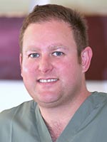 Nick Fahey divides his time between Harley Street Dental Studio in London and Woodborough House Referral Practice in Pangbourne, Berkshire. In both practices he works as a specialist prosthodontist with a special interest in oral surgery and dental implantology. Nick Fahey divides his time between Harley Street Dental Studio in London and Woodborough House Referral Practice in Pangbourne, Berkshire. In both practices he works as a specialist prosthodontist with a special interest in oral surgery and dental implantology.
After training in his native New Zealand, Nick also studied at the Eastman Dental Institute. There he obtained the M.Clin.Dent degree in Prosthodontics. Nick also holds the MRD RCS (Ed), FRACDS and MFDS RCS (Eng).
His areas of interest include all aspects of dentistry related to dental implants and fixed and removable prosthodontics.
Of particular recent interest has been his involvement in digital dentistry. He is particularly interested in computer-guided surgery for simplification of surgical placement of dental implants. This is facilitated by using C.T. scans in conjunction with virtual implant planning software, intraoral scans and 3D printing of surgical guides.
Nick enjoys membership of the ADI and the ITI, where he is a study club director. He has presided over the LDSC and was a founding member of the NZDSL. He also is a past president of the Reading Branch of the BDA (2012).
Qualifications: BDS MClinDent (Pros) FRACDS, MRD RCS (Ed) MFDS RCS (Eng)
|
| 15:50 to 16:10 |
Upper right first molar tooth replacement in the site of previous oro-antral-communication This is the case of a fit and healthy 35 year old female patient who requested the replacement of her missing upper right first molar (UR6).
Approximately three years prior to presentation the UR6 was displaced into the right antrum during a routine extraction. The patient was referred to a Maxillofacial unit to retrieve the displaced tooth and repair the resulting oral antral fistula with a buccal advancement flap.
Clinical and CT scan investigation demonstrated a grossly deficient alveolus in both width and height, and a lack of keratinised mucosa. The prosthetic space was uncompromised and had appropriate mesiodistal and interocclusal space. All fixed and removable prosthodontics options were fully discussed to allow the patient to make an informed decision. Her preference was to replace the UR6 with a single tooth implant.
A two-staged surgical approach was undertaken. The first stage involved a sinus elevation and augmentation with bio-oss with a simultaneous autogenous block graft to restore ridge width. A submerged 4.6mm x 9mm Biohorizons implant was placed at second stage. A coronal advancement flap was performed at the time of exposure to augment the buccal keratinised mucosa. A screw retained metal-ceramic crown was chosen as the definitive restoration, to aid future prosthetic maintenance.
Summary: This case is an example of a severely compromised UR6 edentulous space with respect to both bone and soft tissue. It demonstrates a complex three dimensional bone augmentation, together with soft tissue augmentation to allow for a single tooth implant in the UR6 position.
Paul Swanson 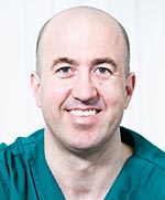 Paul Swanson qualified from the University of Liverpool in 2000. His career has mainly been based in General Dental Practice but has also combined this with part time hospital based oral surgery. He completed his MFGDP qualification in 2003 and a Post Graduate Certificate in Conscious Sedation in 2004. In 2008 he received the Diploma in Implant Dentistry from the Royal College of Surgeons of England and the Advanced Certificate in Bone Grafting. In 2011 he received a Master of Science degree in Implant Dentistry from Queen Mary University of London. Paul Swanson qualified from the University of Liverpool in 2000. His career has mainly been based in General Dental Practice but has also combined this with part time hospital based oral surgery. He completed his MFGDP qualification in 2003 and a Post Graduate Certificate in Conscious Sedation in 2004. In 2008 he received the Diploma in Implant Dentistry from the Royal College of Surgeons of England and the Advanced Certificate in Bone Grafting. In 2011 he received a Master of Science degree in Implant Dentistry from Queen Mary University of London.
Over the last 7 years the volume and complexity of cases has naturally increased. Paul’s clinical practice is now limited to Implant Dentistry. He owns a specialist referral practice in Liverpool and contributes to the multidisciplinary cases done by his team. He mentors dentists at both entry level and advanced implantology and contributes to study clubs in his region.
Qualifications: BDS MFGDPUK PGCert (Sed) DipImpDent RCS (Eng) MSc (Impl) |
| 16:10 to 16:30 |
Soft and hard tissue reconstruction of a severely deficient site prior to implant placement This case report describes how a severe localized alveolar bone deficiency together with altered gingival contour in the site of upper left first molar was reconstructed. A sinus exposure following earlier extraction and an attempt to close it with a buccal advancement flap compromised the site even further by repositioning the buccal fraenum in an unfavourable position.
This case was managed in three stages. First, a sinus lift procedure was performed through the floor of the alveolar defect after extending the existing sinus floor perforation. This, needed minimal bone removal. This space and the alveolar defect was then reconstructed with Regenaform and Bio-guide. Six months afterwards, a free gingival graft was harvested from the left side of the palate and grafted to the UL6 site after raising a full mucoperiosteal flap incorporating the buccal fraenum and repositioning it apically. Two months later, a 13 mm, wide platform Nobel Replace Tapered implant was inserted in the healed site. After three months, a crown was fabricated and fitted.
With this technique, both sinus augmentation and alveolar reconstruction were performed simultaneously using Regenaform which was less invasive to the patient as it was carried out in one surgery. The altered gingival contour was also corrected by performing a free gingival graft on the second stage. This increased the width of keratinized gingiva around the implant which will improve the health of perimplant tissue and improve the gingival contour around UL5.
This case report was published in the 1/2013- Vol. 9 edition of EDI journal.
Younes Khosroshahy 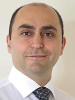 Younes Khosroshahy qualified from Faculty of Dentistry, Tehran Medical University in 1996 with first class honours. Having worked for three years in general dental practice, he moved to the UK in 2000 for postgraduate training in Oral and Maxillofacial Surgery. He achieved the Membership of the Royal College of Surgeons of England in 2003 after working in various London hospitals for three years. Younes Khosroshahy qualified from Faculty of Dentistry, Tehran Medical University in 1996 with first class honours. Having worked for three years in general dental practice, he moved to the UK in 2000 for postgraduate training in Oral and Maxillofacial Surgery. He achieved the Membership of the Royal College of Surgeons of England in 2003 after working in various London hospitals for three years.
Having passed the IQE examination of the GDC in 2004, he then worked as a Staff Grade Oral Surgeon in the Queen Victoria Hospital, East Grinstead and also in a general dental practice in London for four years. During this time, he developed a keen interest in dental implants. He joined Hospital Lane Dental and Implant Clinic in 2007 and has worked there since then. He passed the rigorous examination of the Royal College of Surgeons of Edinburgh and obtained the Diploma in Implant Dentistry in 2010.
Qualifications: DDS MFDS RCS(Eng) Dip Imp Dent RCSEd |
| 16:30 to 17:00 |
Panel Discussion
Forum A - Panel 1
|
| 17:00 |
Close of Forum
|